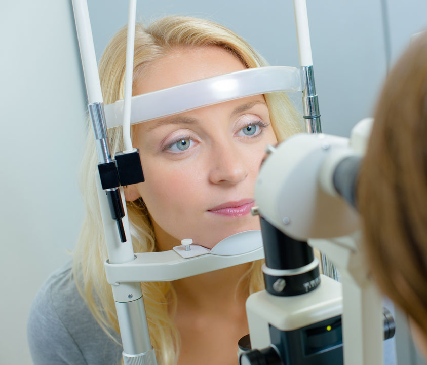
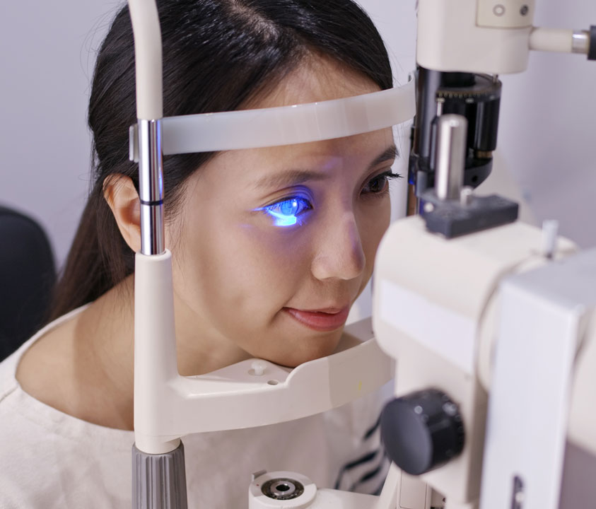
MEDICAL EYE CARE

Many health conditions can cause vision loss. Medical eye exams can help manage and diagnose many diseases.

MEDICAL EYE CARE

Many health conditions can cause vision loss. Medical eye exams can help manage and diagnose many diseases.

medical eye treatments at palm valley eye

Edward Gorak

“EXCELLENT PATIENT EXPERIENCE. DR. SCOTT CARLTON AND HER STAFF ARE CARING, EMPATHETIC, PERSONABLE AND THOUGHTFUL.”

Edward Gorak

“EXCELLENT PATIENT EXPERIENCE. DR. SCOTT CARLTON AND HER STAFF ARE CARING, EMPATHETIC, PERSONABLE AND THOUGHTFUL.”

MEDICAL EYE SERVICES AT PALM VALLEY EYE

Medical Eye Disease can be a devastating problem for many Americans. Fortunately, many of the diseases that commonly cause blindness can be managed when caught early and treated appropriately. An annual comprehensive eye exam is the best way to detect and monitor for the development of blinding eye disorders.

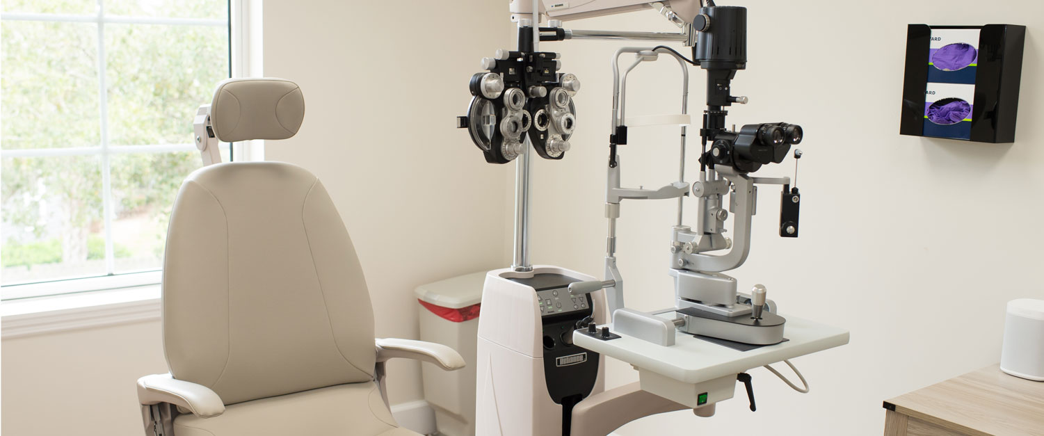
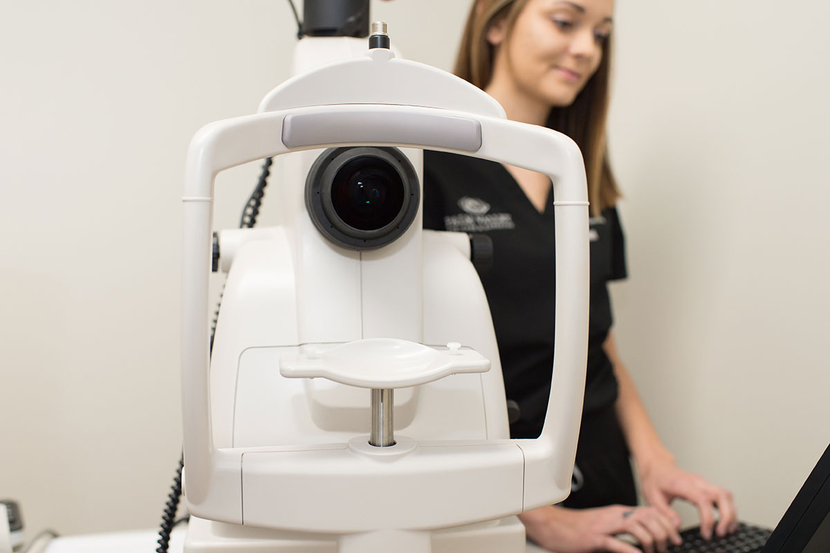
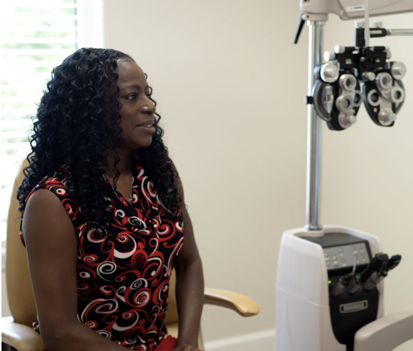
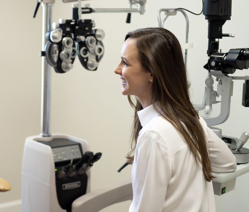
That’s why, at Palm Valley Eye, we do everything we can to make ourselves available to our patients, and to make sure you receive a thorough comprehensive eye exam at least once a year. That’s what compassionate eye care is all about.

Palm Valley Eye Medical Solutions

Of course, step one is a comprehensive eye exam with your doctor to accurately diagnose your medical eye condition in order to discuss and provide the most appropriate treatment for you.

DRY EYE SYNDROME

Dry Eye Syndrome is irritation and inflammation of the eye surface, due to lack of or poor quality tears.

It is a common problem caused by a wide range of conditions.

Symptoms of Dry Eye

People often experience dry eye as they age. Symptoms include foreign body sensation, burning, gritty or sandy sensation, stinging, dryness, eye redness, discomfort in contact lenses, and sensitivity to light.

In some patients, dry eye may be related to an underlying systemic condition, such as rheumatoid arthritis, Sjogrens disease, or medications.

dry eye treatment at palm valley eye care
Dry Eye Syndrome is irritation and inflammation of the eye surface, due to lack of or poor quality tears.

It is a common problem caused by a wide range of conditions.

Symptoms of Dry Eye

People often experience dry eye as they age. Symptoms include foreign body sensation, burning, gritty or sandy sensation, stinging, dryness, eye redness, discomfort in contact lenses, and sensitivity to light.

In some patients, dry eye may be related to an underlying systemic condition, such as rheumatoid arthritis, Sjogrens disease, or medications.

Treatment of Dry Eye

For most patients, mild symptoms can be managed with over-the-counter lubricants and attention to environmental influences. More serious and persistent symptoms require evaluation for underlying conditions. Treatments in these cases vary and are best discussed with your doctor.

In many cases simple lifestyle changes can help including:

• Avoiding environmental irritants
• Using recommended over-the-counter lubricating drops regularly
• Attention to lid margin and oil gland hygiene
• Eliminating medications that may be responsible
• Adding Omega-3 fatty acids to your diet
• Taking supplements recommended by your doctor

If these methods are unsuccessful, the following treatments may be an option:

• Prescription eye drops
• Prescription oral medications
• Insertion of punctal plugs to limit tear drainage
• Punctal cautery to permanently close tear drainage
• Amniotic membrane placement
• Surgical correction of eyelid anatomy or tarsorrhaphy in severe cases

EYE IRRITATION

Eye irritation is a daily problem for many people with symptoms ranging from redness, to itching or swelling. Allergies are a likely culprit, but eye irritations may also be caused by non-emergency conditions or in rare cases, dangerous conditions. Any irritation that is persistent should be treated with direction from an eye specialist.

GLAUCOMA

Most people with glaucoma have no early symptoms. The disease slowly reduces peripheral vision and can go unnoticed until significant damage has occurred. Risk factors for glaucoma include increased age, family history, African American, Hispanic, or Asian heritage, chronic use of steroids, and high eye pressures.

The damage caused by glaucoma cannot be reversed. Regular checkups and appropriate treatment are the best methods to slow or prevent vision loss, especially if caught in the early stages.

What is Glaucoma?

Glaucoma is the deterioration of the optic nerve as a result of abnormal eye pressure. Studies show that other factors likely contribute to disease, which is why some patients have “normal” pressure glaucoma.

Symptoms of Glaucoma

• Often Asymptomatic
• Peripheral Vision Loss
• Painless

How We Test for Glaucoma

Glaucoma testing includes intraocular pressure measurement, measurement of your corneal thickness, clinical examination of your ocular structures and optic nerve, and formal testing with visual fields and nerve fiber layer OCT imaging. At Palm Valley Eye, we generally spread these tests out over separate visits to make the examination more comfortable.

Glaucoma Treatments

Glaucoma Treatments

There is no cure for glaucoma, but the disease can be stabilized and vision loss prevented if caught early.

Treatments for glaucoma include prescription eye drops, laser treatment, minimally invasive surgical implants, and surgery with a drainage device or trabeculectomy.

Laser Iridotomy
Laser Peripheral Iridotomy is the standard first-line treatment in closed angle glaucoma and eyes at risk for this condition.

The “angle” is the space between the clear part of the eye (cornea), and the colored part (iris). It contacts the main structure that directs fluid out of the eye.

In closed angle glaucoma, the angle is closed in many or most areas, causing increased eye pressure, optic nerve damage and possible vision loss. The laser creates a hole in the outer edge of the iris, which widens (opens) the angle and enhances fluid outflow.

While this procedure will not improve your vision, it is intended to preserve vision and prevent glaucoma from progressing.

Selective Laser Trabeculoplasty
SLT is a form of laser surgery that is used to lower the intraocular pressure in Glaucoma. It is FDA approved.

Laser energy is applied to the drainage structure in the angle of the eye. This causes a chemical and biological change in the tissue that results in better drainage of fluids out of the eye, and a lowering of the pressure in a large number of patients.

SLT lowers the intraocular pressure by about 30% in responsive patients. Your doctor may have you continue your regular glaucoma drops after the procedure. SLT may be performed more than once and generally is effective for several years. SLT is considered safe and is performed as an outpatient procedure.

While this procedure will not improve your vision, it is intended to preserve vision and prevent glaucoma from progressing.

MACULAR DEGENERATION

Age-related macular degeneration (AMD/ARMD) is the leading cause of low vision over the age of 65. The largest risk factor for macular degeneration is age. If you are 55 or older, we strongly encourage regular dilated examinations to detect early signs of ARMD. Other important risk factors are family history and smoking.

In early stages, vision may not be affected. Progression of the disease may lead to blurred or distorted central vision or eventual permanent vision loss. Urgent treatment with vision changes is imperative to prevent permanent vision loss. In some stages of disease, progression may be slowed by vitamin use.

Specifics of Macular Degeneration

Macular Degeneration is caused by the deterioration of the central portion of the retina, known as the macula. The macula is responsible for central vision and controls our ability to read, drive a car, recognize faces and see objects in detail.

Patients with ARMD may experience partial vision loss, blurred vision, distorted vision, or see waves and spots in their vision. It is painless and may worsen slowly over time or suddenly cause vision changes.

ARMD is currently considered an incurable disease, but if caught early through regular eye exams, the risks can be reduced or the progression slowed.

macular degeneration treatments at palm valley eye
Age-related macular degeneration (AMD/ARMD) is the leading cause of low vision over the age of 65. The largest risk factor for macular degeneration is age. If you are 55 or older, we strongly encourage regular dilated examinations to detect early signs of ARMD. Other important risk factors are family history and smoking.

In early stages, vision may not be affected. Progression of the disease may lead to blurred or distorted central vision or eventual permanent vision loss. Urgent treatment with vision changes is imperative to prevent permanent vision loss. In some stages of disease, progression may be slowed by vitamin use.

Specifics of Macular Degeneration

Macular Degeneration is caused by the deterioration of the central portion of the retina, known as the macula. The macula is responsible for central vision and controls our ability to read, drive a car, recognize faces and see objects in detail.

Patients with ARMD may experience partial vision loss, blurred vision, distorted vision, or see waves and spots in their vision. It is painless and may worsen slowly over time or suddenly cause vision changes.

ARMD is currently considered an incurable disease, but if caught early through regular eye exams, the risks can be reduced or the progression slowed.

Macular Degeneration Treatments

Macular Degeneration Treatments

Treatments for Early Dry ARMD
The treatment for early dry ARMD is generally nutritional therapy, with a healthy diet high in antioxidants to support the cells of the macula and in some cases, prescription vitamins known as AREDS-2. We also strongly encourage smoking cessation and are available to assist you in this goal.

An important part in this stage of disease is close monitoring for new vision changes, which may signify conversion to “wet” ARMD. This is done most commonly by using an Amsler grid.

Treatments for Wet ARMD
Wet, or exudative, ARMD is treated with intraocular injections in most cases. The risk of permanent vision loss increases with delayed treatment so we strongly encourage any patient with ARMD and vision changes to contact their doctor today.

EYE PAIN

Eye Pain takes many forms and can occur in all areas of the eye. The pain can be persistent or random; sharp or dull. Eye pain that does not subside after rest with over the counter medication may be caused by something serious and should be presented to an eye specialist as soon as possible for diagnosis.

ALLERGIES

Eye allergies are no different than allergies that affect your sinuses, nose or lungs. Common offenders include seasonal pollen, dust, pets, and medications.

Symptoms: Itching, Tearing, Redness, Foreign body sensation

Treatments: Avoid exposure to known airborne allergens, such as pollen. Avoid rubbing your eyes. Topical lubricating drops, oral allergy medications, and topical prescription drops may be used.

AMBLYOPIA

Amblyopia is a term used for an eye that does not see well due to abnormal development of sight during early childhood. When one eye develops good vision while the other does not, the eye with poorer vision is called amblyopic. Amblyopia typically involves one eye, but can involve both eyes on occasion.

The best time to correct amblyopia is during infancy or early childhood. The American Academy of Pediatric Ophthalmology and American Academy of Pediatrics recommend screening as a newborn in the hospital, by 6 months with their pediatrician well visits, use of photo screening devices with the pediatrician well visits starting at ages 1-2, and again at ages 3-4.

After 5 years, the child should have regular vision checked in both eyes during their pediatric visits. Often the primary care doctor will refer the child to an ophthalmologist for any abnormalities or screening concerns.

Treatment of amblyopia is most successful when caught early. The goal of treatment is to force the child to use the weak eye. Treatments often include prescription glasses, patching, use of eye drops, and occasionally surgery to correct any muscle (strabismus) abnormalities.

BLEPHARITIS

Blepharitis is a chronic or long-term inflammation of the eyelids. It affects people of all ages. Blepharitis is often caused by poor eyelid hygiene resulting in clogged oil glands located in the eyelid margins, bacterial or demodex infection, or an allergic reaction. Blepharitis commonly causes chalazions and styes.

Some patients with systemic conditions like rosacea are more prone to developing blepharitis.

Treatment of blepharitis includes improved eyelid hygiene using warm compresses and lid scrubs routinely. Additional treatments may include use of prescription drops and ointments, oral medications, and manual expression of glands in the office.

CONJUNCTIVITIS (“PINK EYE”)

Conjunctivitis is irritation and inflammation of the conjunctiva – this is the clear layer overlying the sclera, or white part of the eye. Conjunctivitis is commonly caused by allergies, irritants, and viral infections. Rarely, it may be caused by a bacterial infection.

Symptoms commonly include eye redness, eyelid swelling, crusting or sticking of the lids together, rope-like discharge, tearing, itching, burning, light sensitivity, and foreign body sensation. Usually both eyes are involved, although symptoms may start in one eye and go to the other, or one eye may be worse than the other.
Treatment of conjunctivitis varies based on the underlying condition.

– Seasonal allergies can be treated with oral allergy medication, lubricating drops, and topical allergy drops.

– In conjunction with a runny nose, cough, or congestion, a viral cause is most likely and it is important to use appropriate hygiene to avoid spreading to others. Viral conjunctivitis is highly contagious and you should avoid contact with others, wash your hands with soap regularly, avoid touching your face and eyes, throw away contaminated cosmetic products, and avoid reuse of towels and pillow cases. Antibiotics do not treat viruses, but patients can obtain comfort through use of cold lubricating drops, cold compresses, and occasionally prescription drops from their eye doctor.

– Bacterial conjunctivitis is a rare, but serious condition. Usually only one eye is involved and symptoms of eye swelling, lid swelling, and redness may be severe. Thick white or yellow discharge (purulent) is a feature. Bacterial conjunctivitis requires evaluation and treatment by your ophthalmologist with topical prescription drops and occasionally oral medications.

macular degeneration treatments at palm valley eye
Conjunctivitis is irritation and inflammation of the conjunctiva – this is the clear layer overlying the sclera, or white part of the eye. Conjunctivitis is commonly caused by allergies, irritants, and viral infections. Rarely, it may be caused by a bacterial infection.

Symptoms commonly include eye redness, eyelid swelling, crusting or sticking of the lids together, rope-like discharge, tearing, itching, burning, light sensitivity, and foreign body sensation. Usually both eyes are involved, although symptoms may start in one eye and go to the other, or one eye may be worse than the other.
Treatment of conjunctivitis varies based on the underlying condition.

– Seasonal allergies can be treated with oral allergy medication, lubricating drops, and topical allergy drops.

– In conjunction with a runny nose, cough, or congestion, a viral cause is most likely and it is important to use appropriate hygiene to avoid spreading to others. Viral conjunctivitis is highly contagious and you should avoid contact with others, wash your hands with soap regularly, avoid touching your face and eyes, throw away contaminated cosmetic products, and avoid reuse of towels and pillow cases. Antibiotics do not treat viruses, but patients can obtain comfort through use of cold lubricating drops, cold compresses, and occasionally prescription drops from their eye doctor.

– Bacterial conjunctivitis is a rare, but serious condition. Usually only one eye is involved and symptoms of eye swelling, lid swelling, and redness may be severe. Thick white or yellow discharge (purulent) is a feature. Bacterial conjunctivitis requires evaluation and treatment by your ophthalmologist with topical prescription drops and occasionally oral medications.

CORNEAL DYSTROPHY

There are many forms of corneal dystrophies. Two of the more common types are Anterior Basement Membrane Dystrophy (also known as Map Dot Dystrophy or Epithelial Basement Membrane Dystrophy) and Fuch’s Endothelial Dystrophy.

Anterior Basement Membrane Dystrophy is a disorder that causes the cells on the very top, or front part of the eye, (the epithelium) to stick together poorly. Patients often complain of waking up with eye pain or scratches. ABMD can also cause frequent changes in glasses prescriptions and dry eye symptoms. Treatment varies based on the severity and typically involves use of lubricating drops, prescription drops, contact lenses, and occasionally surgical or laser treatments.

Fuch’s Endothelial Dystrophy is a disorder that causes the cells on the back part, or inside part, of the cornea (the endothelium) to function poorly and develop abnormal structures called guttae. These cells typically pump fluid out of the cornea, so when they don’t work well, the cornea swells leading to blurry vision. Guttae can also cause symptoms of glare or changes in the quality of vision. This condition is often treated with topical drops when mild. Severe disease can effectively be treated with a partial corneal transplant known as endothelial keratoplasty (DSEK and DMEK).

CORNEAL INFECTION OR ULCER

Corneal ulcers are infections of the clear part of the eye. They are extremely painful, cause redness of the eye, sensitivity of light, and often headache. They are commonly associated with improper contact lens wear, although a scratch or abrasion of the surface can also lead to infection. Other less common causes include extreme surface dryness or exposure keratopathy and herpetic infections.
Corneal infections and ulcers are dangerous conditions and require urgent evaluation with your ophthalmologist. Several drops are often required for treatment, which depends on the type of infection. Without treatment, infection of the cornea can lead to scarring, vision loss, infection inside the eye or around the eye, and even spread of infection to the brain.

DIABETIC RETINOPATHY

Diabetic Retinopathy is the most common cause of vision loss in working aged adults. If you are diabetic, it is essential that you have yearly dilated examinations with a professional.

Diabetes leads to damaged blood vessels in the retina and can cause bleeding, loss of oxygen to the retina tissue (cotton wool spots), build up of waste products (drusen and exudates), and swelling of the retina (macular edema). In the early stages, known as non-proliferative diabetic retinopathy, changes can be reversed with good control of blood sugar, blood pressure, and cholesterol.

In advanced stages, known as proliferative diabetic retinopathy, dangerous blood vessels grow from the retina and cause bleeding into the eye, growth of scar tissue, neovascular glaucoma, and traction retinal detachments. These changes often lead to irreversible blindness. The severe stage of diabetic retinopathy requires treatment with lasers, injections, and often surgery. The best way to prevent vision loss from diabetic retinopathy is to ensure early detection through regular dilated examinations and maintain good control of your blood sugar, blood pressure, and cholesterol.

FLASHES AND FLOATERS

Flashes and floaters may be associated with several eye conditions including posterior vitreous detachment, retinal breaks and tears, retinal detachments, or bleeding inside the eye. If you are experiencing these symptoms, it is important to be evaluated by your ophthalmologist urgently.

RETINAL TEAR OR DETACHMENT

Retinal tears are breaks in the lining of the back part of the eye, or retina. These are commonly caused by detachment of the gel part of the eye from the retina during which the mechanical tugging causes a break. Retinal tears, if not treated promptly, can lead to retinal detachments which occur from fluid traveling through the retinal break and lifting the retina from the back part of the eye. This is a serious condition and can lead to permanent vision loss, especially when not treated promptly.

Symptoms of flashes, floaters, appearance of a curtain, or loss of parts of the vision could all be signs of a retinal tear or detachment and require urgent evaluation with your ophthalmologist.

Patients with a history of high myopia (near sightedness), lattice degeneration, prior eye surgery, prior history of retinal tears/detachment, prior eye injury or family history are at increased risk.

Retinal tears and breaks are treated with laser, and occasionally injection of a gas bubble into the eye. Retinal detachments often require surgical repair.

retinal tear at palm valley eye
Retinal tears are breaks in the lining of the back part of the eye, or retina. These are commonly caused by detachment of the gel part of the eye from the retina during which the mechanical tugging causes a break. Retinal tears, if not treated promptly, can lead to retinal detachments which occur from fluid traveling through the retinal break and lifting the retina from the back part of the eye. This is a serious condition and can lead to permanent vision loss, especially when not treated promptly.

Symptoms of flashes, floaters, appearance of a curtain, or loss of parts of the vision could all be signs of a retinal tear or detachment and require urgent evaluation with your ophthalmologist.

Patients with a history of high myopia (near sightedness), lattice degeneration, prior eye surgery, prior history of retinal tears/detachment, prior eye injury or family history are at increased risk.

Retinal tears and breaks are treated with laser, and occasionally injection of a gas bubble into the eye. Retinal detachments often require surgical repair.

HALOS

Halos appear as a fuzzy ring around lights. Patients often complain of these when faced with headlights while driving at night. Most halos are associated with cataracts, but other refractive changes such as astigmatism or corneal changes may cause them. Rarely, halos can be seen with dangerous eye conditions. If you experience eye pain, headache, nausea, or change in vision with your halos contact your ophthalmologist immediately.

HERPES SIMPLEX KERATITIS

Herpes Simplex is a viral infection that lives in the nerve roots and can reactivate to involve the skin and eyes. A prior history of cold sores is common. Signs and symptoms include a skin rash around the eyes (painful blisters), eye redness, irritation, burning, tearing, foreign body sensation, light sensitivity, and pain. Sometimes ocular HSV infection does not present with a skin rash and instead causes what are thought to be recurrent or repetitive scratches of the eye. This is an urgent condition and you should be seen by an ophthalmologist for appropriate evaluation and treatment.

Treatment of herpes keratitis involves oral and topical prescription medications. Because this disease can recur, it is important to be followed by an ophthalmologist regularly to avoid corneal scarring and loss of vision.

IRITIS (UVEITIS)

Iritis (a form of uveitis) is inflammation of the front part of the eye. Symptoms include eye redness, pain, sensitivity to light, and decreased vision. Iritis is commonly caused by blunt trauma or injury. Most cases not related to trauma are idiopathic (meaning no known underlying cause). Recurrent episodes are often related to systemic disease.

Iritis or uveitis is a serious condition and requires evaluation and treatment with your ophthalmologist. Without treatment, inflammation in the eye can lead to scarring, vision loss, glaucoma, and blindness.

KERATOCONUS

Keratoconus is a corneal dystrophy that causes abnormal thinning and an atypical cone shape of the cornea. This often presents in middle-aged adults with frequent prescription changes, decreased vision, and irregular astigmatism. Patients may require hard contact lenses, or rigid gas permeable lenses, to obtain their best vision. Other symptoms of keratoconus include episodes of eye pain, redness, and blurry vision associated with corneal hydrops, or acute breaks in the barrier layer of the cornea that lead to swelling and eventual scarring.

There are many ways to manage keratoconus including use of glasses, soft contact lenses, and hard contact lenses. In the early stages of keratoconus, patients may be eligible for corneal cross linking to slow progression of the changes in corneal shape. Episodes of hydrops are often treated with topical eye drops and occasionally use of an air bubble inside the eye. When patients have severe disease or scarring, corneal transplant surgery may be needed.

medical eye care exam at palm valley eye care
Keratoconus is a corneal dystrophy that causes abnormal thinning and an atypical cone shape of the cornea. This often presents in middle-aged adults with frequent prescription changes, decreased vision, and irregular astigmatism. Patients may require hard contact lenses, or rigid gas permeable lenses, to obtain their best vision. Other symptoms of keratoconus include episodes of eye pain, redness, and blurry vision associated with corneal hydrops, or acute breaks in the barrier layer of the cornea that lead to swelling and eventual scarring.

There are many ways to manage keratoconus including use of glasses, soft contact lenses, and hard contact lenses. In the early stages of keratoconus, patients may be eligible for corneal cross linking to slow progression of the changes in corneal shape. Episodes of hydrops are often treated with topical eye drops and occasionally use of an air bubble inside the eye. When patients have severe disease or scarring, corneal transplant surgery may be needed.

PINGUECULA

Pingueculas are discolored changes on the front part of the eye associated with sun damage. They commonly appear as yellow spots on the white part of the eye. Usually pingueculas are asymptomatic and not dangerous. Topical lubricating drops may be used for discomfort and sunglasses are recommended for UV protection. Rarely, these spots may become inflamed and require evaluation and treatment with your eye doctor.

PTERYGIUM

Pterygiums are discolored changes on the front part of the eye associated with sun damage. They commonly appear as white and red growths from the inside part of the eye onto the clear part, or cornea. Pterygiums are often asymptomatic in the early stages, but may grow and causes changes or decrease in vision. Treatment of pterygiums include topical lubricating drops, topical prescription drops for inflammation or irritation, and surgical removal when necessary. Sunglasses are recommended for UV protection.

PRESBYOPIA

Presbyopia is a normal aging phenomenon in which your ability to see up close deteriorates. This is caused by hardening of the lens structure which becomes stiff and unable to change shape to pull things in focus. Difficulty reading is best addressed with use of reading glasses, bifocals, or monovision techniques. With some of the newer cataract surgery technology, some patients may benefit from multifocal intraocular lenses.

Occasionally, patients may note over time that they have developed the ability to read again, or as we call it “second sight”. This is typically related to the progressive increase in the lens density or cataract, which is a refractive phenomenon, and associated with decreased distance vision.

LARGE OR SMALL PUPIL

The pupil is the circular opening in the colored part of the eye, seen as a black circle. Pupils change in size depending on the amount of light entering the eye. In dark conditions the pupil is larger, while in bright conditions the pupil becomes smaller. Most people have pupils that are about the same size in both eyes. New changes in the size of one pupil can be associated with exposure to medications or chemicals or other systemic conditions and should be evaluated by your eye doctor.

DROOPING EYELID (PTOSIS)

A drooping eyelid, formally called Ptosis, is when the upper eyelid is lower in position than typical. This is most commonly a result of changes in the connective tissues of the eyelid caused by aging or trauma. However, if this has occurred suddenly or is associated with other symptoms, you should be evaluated by your ophthalmologist urgently.

SHINGLES (HERPES ZOSTER)

Shingles, or Herpes Zoster, is a viral infection that lives in the nerve roots and can reactivate to involve the skin and eyes. It is the same virus that causes chicken pox. Signs and symptoms include a skin rash on one side of the face (very painful blisters and scabs), eyelid redness and swelling, eye redness, irritation, burning, tearing, foreign body sensation, light sensitivity, and pain. Sometimes the ocular infection does not present with a skin rash and instead causes eye symptoms or vision changes. Recurrent episodes of this disease can cause severe corneal scarring and vision loss. In some patients, involvement of the retina can lead to blindness. This is an urgent condition and you should be seen by an ophthalmologist for appropriate evaluation and treatment.

Treatment of herpes zoster involves oral and topical prescription medications. Because this disease can recur, it is important to be followed by an ophthalmologist regularly to avoid corneal scarring and loss of vision.

medical eye care shingles
Shingles, or Herpes Zoster, is a viral infection that lives in the nerve roots and can reactivate to involve the skin and eyes. It is the same virus that causes chicken pox. Signs and symptoms include a skin rash on one side of the face (very painful blisters and scabs), eyelid redness and swelling, eye redness, irritation, burning, tearing, foreign body sensation, light sensitivity, and pain. Sometimes the ocular infection does not present with a skin rash and instead causes eye symptoms or vision changes. Recurrent episodes of this disease can cause severe corneal scarring and vision loss. In some patients, involvement of the retina can lead to blindness. This is an urgent condition and you should be seen by an ophthalmologist for appropriate evaluation and treatment.

Treatment of herpes zoster involves oral and topical prescription medications. Because this disease can recur, it is important to be followed by an ophthalmologist regularly to avoid corneal scarring and loss of vision.

STYE

A stye is inflammation and/or infection of the oil glands on the eyelid. It is often painful and appears as a red bump near an eyelash. Recurrent styes are often caused by an underlying condition called blepharitis. Treatment of styes includes warm compresses, gently lid scrubs and massages, and lubricating drops. Rarely, topical or oral medications may be needed.

Improve Your Vision at Palm Valley Eye Care & Surgeons.

Fill out our request appointment form to find out how we can help or call our clinic to learn more.
Most major insurances are accepted.

Appointment: 904-712-3315

Clinic Location:
Ponte Vedra Beach